非洲爪蟾(Xenopus laevis)胚胎易于获取、尺寸大(直径约 1.2 mm)、发育快速且与高等脊椎动物高度同源,可在简单盐溶液中体外完成从受精到蝌蚪期的全程发育,是研究脊椎动物早期发育的经典模式系统。Xenopus laevis embryos are large (~1.2 mm), rapidly developing and highly conserved with higher vertebrates. They develop in vitro in simple saline from fertilisation to tadpole stage — a classic model for early vertebrate development.
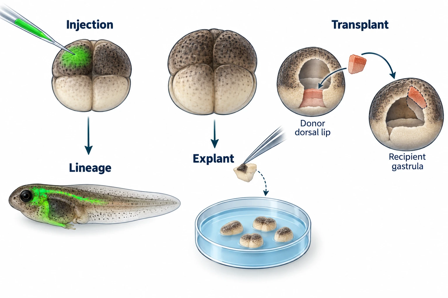
核心优势Core advantages
- 胚胎可及性:hCG 诱导排卵可大量获取同步发育胚胎,全年均可操作,供应稳定。Embryo accessibility: hCG-induced ovulation provides large numbers of synchronously developing embryos year-round.
- 操作可视性:胚胎直径约 1.2 mm,动物极(深色)与植物极(淡黄色)分化清晰,在解剖镜下即可精确定位并完成显微注射,无需荧光引导。Visibility: ~1.2 mm diameter with clearly distinct animal (dark) and vegetal (pale) poles — precise microinjection without fluorescence guidance.
- 体外发育:在 0.1× MBS-H 等简单盐溶液中可完成受精至蝌蚪期的全程发育,方便实时观察与干预。In vitro development: complete development from fertilisation to tadpole in simple saline (0.1× MBS-H); easy to observe and manipulate at any stage.
- 遗传工具兼容:支持 mRNA 过表达、Morpholino 反义寡核苷酸(翻译阻断或剪接干扰)与 CRISPR-Cas9 基因编辑,可灵活组合功能获得与功能丧失实验。Genetic tool compatibility: compatible with mRNA overexpression, Morpholino knockdown (translation block or splice switching) and CRISPR-Cas9 — gain-of-function and loss-of-function flexibly combined.
主要研究方法Key methods
1. 胚胎获得与显微操作Embryo collection & micromanipulation
- hCG 注射诱导排卵(雌蛙 500 IU),获取同步受精卵;4% L-半胱氨酸(pH 7.8–8.0)去除卵胶膜后进行操作。hCG injection (female 500 IU) for synchronised ovulation; remove jelly coat with 4% L-cysteine (pH 7.8–8.0) before manipulation.
- 拉制 1–5 μm 尖端的玻璃针,校准注射体积(1–20 nL/次),可靶向特定卵裂球,精确导入 mRNA、蛋白质或 Morpholino。Pull glass needles (1–5 μm tip), calibrate volume (1–20 nL per injection); target specific blastomeres for precise delivery of mRNA, protein or Morpholino.
2. 细胞谱系追踪Cell lineage tracing
- 将荧光染料(DiI、荧光葡聚糖 Fluorescent Dextran)或 GFP mRNA 显微注射至目标区域的单个或一群细胞。Microinject fluorescent dyes (DiI, fluorescent dextran) or GFP mRNA into target cells.
- 在共聚焦显微镜下实时追踪细胞迁移轨迹、克隆扩增与命运图谱,分辨率可至单细胞水平;结合延时成像可记录完整的发育动态。Track cell migration, clonal expansion and fate map in real time by confocal microscopy at single-cell resolution; time-lapse imaging captures full developmental dynamics.
3. 胚胎切割与移植(Spemann 组织者实验)Embryo cutting & grafting (Spemann organizer assay)
- 将背唇(Spemann 组织者)切割后移植至宿主胚胎腹侧,可诱导宿主腹侧形成第二套完整体轴——经典实验验证了组织者的诱导能力与跨物种保守性。Graft the dorsal lip (Spemann organizer) onto the ventral side of a host embryo — induces a complete second body axis, demonstrating organizer inductive activity and cross-species conservation.
- 外植块(Explant)培养:切取动物帽(Animal cap)或其他区域组织体外培养,分离信号通路的直接诱导效应,排除体内间接影响。Explant culture: excise animal cap or other tissue regions for in vitro culture, isolating direct signalling effects and excluding indirect in vivo influences.
4. 基因功能研究Gene function analysis
- 功能获得:体外转录目标基因 mRNA,显微注射至单细胞或特定卵裂球,快速分析过表达表型;可精确定时与定位。Gain-of-function: inject in vitro-transcribed mRNA into one-cell or specific blastomeres; fast phenotypic readout with precise temporal and spatial control.
- 功能丧失:Morpholino 阻断翻译或干扰剪接;CRISPR-Cas9 注射引入靶向突变(F0 镶嵌体)或建立稳定遗传系。Loss-of-function: Morpholino for translation block or splice switching; CRISPR-Cas9 for targeted mutagenesis (F0 mosaic) or stable line generation.
- 拯救实验:同时注射 MO 与抗敲低 mRNA,观察表型能否恢复——特异性验证的金标准。Rescue: co-inject MO and MO-resistant mRNA; phenotype rescue is the gold-standard specificity assay.
5. 信号通路解析Signalling pathway analysis
- 信号分子局部微量施加(植入蛋白包被小珠),精确检测特定组织的感受性。Local signal application (protein-coated beads) to assess tissue competence at precise locations.
- 报告基因(GFP/荧光素酶)实时监测启动子活性与信号应答。Reporter genes (GFP/luciferase) for real-time monitoring of promoter activity and signalling responses.
- 原位杂交(ISH)绘制基因时空表达图谱;免疫组化(IHC)定位蛋白分布与修饰状态。In situ hybridisation (ISH) for spatiotemporal expression mapping; immunohistochemistry (IHC) for protein localisation and modification status.
将显微操作、分子遗传干预与表型分析相结合,可在活体三维背景下高效解析脊椎动物早期发育过程。爪蟾胚胎的体外操作简便性与基因工具的丰富性,使其成为发育生物学不可替代的模式生物。Combining micromanipulation, molecular genetics and phenotypic analysis enables efficient dissection of early vertebrate development in a live 3D context. The ease of in vitro manipulation and breadth of genetic tools make Xenopus embryos an irreplaceable developmental biology model.
参考文献References
- Harland R & Gerhart J (1997) Formation and function of Spemann's organizer. Annu Rev Cell Dev Biol 13:611–667.
- Sive HL, Grainger RM, Harland RM (2000) Early Development of Xenopus laevis: A Laboratory Manual. Cold Spring Harbor Laboratory Press.
- Blum M, et al. (2015) Xenopus, an undervalued model organism. Genesis 53(2):1–19.